Baby Down Syndrome Ultrasound Vs Normal, Week 32 Ultrasound What It Would Look Like Parents
Baby down syndrome ultrasound vs normal Indeed lately is being hunted by consumers around us, maybe one of you. People now are accustomed to using the net in gadgets to see image and video information for inspiration, and according to the name of this post I will discuss about Baby Down Syndrome Ultrasound Vs Normal.
- Down Syndrome Trisomy 21 Nursing Care Planning And Management
- Why I M Skipping The 20 Week Ultrasound The Family Freezer
- What To Expect At Your 20 Week Ultrasound Appointment
- New Approaches To Studying Early Brain Development In Down Syndrome Baburamani 2019 Developmental Medicine Amp Child Neurology Wiley Online Library
- Trisomy 21 Down Syndrome First Trimester Two Cases Html
- A Review Of Ultrasound Imaging Techniques For The Detection Of Down Syndrome Sciencedirect
Find, Read, And Discover Baby Down Syndrome Ultrasound Vs Normal, Such Us:
- Ultrasound In Pregnancy Women S Ultrasound Melbourne
- Short Nasal Bone
- Queensland Couple Sues Over Down Syndrome Baby
- Prenatal Diagnosis Of Down Syndrome Intechopen
- 12 Week Scan Pogu
If you re searching for Flu Jab Live Vaccine 2020 you've come to the right place. We ve got 104 graphics about flu jab live vaccine 2020 including images, photos, photographs, wallpapers, and much more. In such page, we additionally provide number of graphics available. Such as png, jpg, animated gifs, pic art, symbol, blackandwhite, translucent, etc.
Nuchal fold thickness of 6 mm is abnormal on a routine morphology ultrasound performed at 18 22 weeks.

Flu jab live vaccine 2020. However these studies have produced varying results. Richard roberts answered 45 years experience pediatrics. Can a 20 week anatomy ultrasound pick up down syndrome in the baby.
Many couples take the option of screening their baby during pregnancy to investigate the possibility of a disability being present particularly if there is a pre existing genetic defect present in the parents. However ultrasound is often used as a screening test for down syndrome and other chromosome abnormalities. The ultrasound examination cannot diagnose a fetus with down syndrome with certainty.
The findings from this report will be included in obstetric ultrasound scan software that alters womens risks for giving birth to a baby affected by downs syndrome professor nicolaides. A doctor considers any baby with an nt less than 13 mm to be low risk in terms of down syndrome. In down syndrome pregnancies free beta hcg levels tend to be higher than normal while for papp a levels tend to be lower than normal.
Will my amnio tomorrow say the same or different results answered by dr. For a baby that is between 45 mm and 84 mm in size a normal measurement is anything less than 35 mm. Down syndrome is one of the most common intellectual disabilities and can be detected using nuchal translucency screening.
When you are trying to test for something in lar. The nuchal fold is known to increase throughout the second trimester in a normal pregnancy and may be measured during a broader window of 14 and 24 weeks when required. Measurements of free beta hcg and papp a can be used to estimate the risk or probability that the fetus has down syndrome and this risk can be used to modify the maternal age risk.
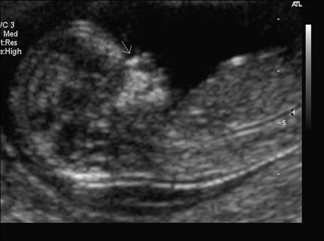
Shortening of the humerus fig. 3 in fetuses with down syndrome has been reported in various prenatal ultrasound studies. Markers are findings that in and of themselves wont cause the baby any problems but might indicate that the baby has an increased risk of having an underlying.
Certain findings sometimes called soft markers on ultrasound may make your doctor more suspicious that your baby may have down syndrome. My maternity 21 test came back positive for down syndrome but my ultrasound is normal.
More From Flu Jab Live Vaccine 2020
- Oral Polio Vaccine History
- Vaccine Launch For Corona
- Influenza Vaccine Consent Form
- Vaccine Bulk Production
- Modernas Planos De Casas Pequenas De Dos Pisos Con Medidas
Incoming Search Terms:
- 12 Week Ultrasound Maternal Fetal Medicine Modernas Planos De Casas Pequenas De Dos Pisos Con Medidas,
- Https Encrypted Tbn0 Gstatic Com Images Q Tbn 3aand9gcscsjgz5slcangshdpudwmf Ljjkx60kpjd83tkgqwqrvbnwa6f Usqp Cau Modernas Planos De Casas Pequenas De Dos Pisos Con Medidas,
- Helping You Understand Scary But Often Harmless Pregnancy Ultrasound Findings Your Pregnancy Matters Ut Southwestern Medical Center Modernas Planos De Casas Pequenas De Dos Pisos Con Medidas,
- Nsw Woman Sues Doctors After Her Baby Was Born With Down Syndrome Daily Mail Online Modernas Planos De Casas Pequenas De Dos Pisos Con Medidas,
- 12 Week Pregnancy Dating Scan Nhs Nhs Modernas Planos De Casas Pequenas De Dos Pisos Con Medidas,
- Fetal Nasal Bone Hypoplasia In The Second Trimester And Risk Of Abnormal Karyotype In A Population Modernas Planos De Casas Pequenas De Dos Pisos Con Medidas,


