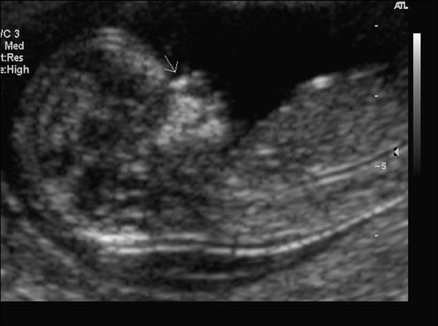
Down Syndrome Ultrasound Vs Normal 13 Weeks, Measuring Nuchal Translucency And Crown Rump Length 12 13 Weeks Scan Youtube
Down syndrome ultrasound vs normal 13 weeks Indeed lately has been hunted by users around us, perhaps one of you personally. Individuals are now accustomed to using the net in gadgets to see image and video data for inspiration, and according to the title of the article I will talk about about Down Syndrome Ultrasound Vs Normal 13 Weeks.
- Nuchal Scan First Trimester Screening Results Normal Range Abnormal Nasal Bone Measurement Baby S Gender
- Genetic Ultrasound Following 1st Trim Screening
- Down Syndrome Markers What Do They Mean For My Baby If They Re Picked Up In An Ultrasound
- Diagnostic Obstetric Ultrasound Glowm
- Nuchal Translucency Nt Normal Range New Health Advisor
- Nuchal Translucency Scan 11 Weeks 14 Weeks Youtube
Find, Read, And Discover Down Syndrome Ultrasound Vs Normal 13 Weeks, Such Us:
- Week 13 Ultrasound What It Would Look Like Parents
- Nuchal Translucency Nt Normal Range New Health Advisor
- Nuchal Scan First Trimester Screening Results Normal Range Abnormal Nasal Bone Measurement Baby S Gender
- Women S Health And Education Center Whec Diagnostic Ultrasound Sonographic Screening For Down Syndrome
- 12 Week Scan So Gi Scan
If you re looking for Pfizer Covid Vaccine Trial Design you've come to the perfect place. We have 104 images about pfizer covid vaccine trial design including images, pictures, photos, wallpapers, and more. In such page, we also provide number of images available. Such as png, jpg, animated gifs, pic art, logo, black and white, translucent, etc.

The Detection Of Spina Bifida At 11 13 6 Weeks Gestation Borg 2017 Sonography Wiley Online Library Pfizer Covid Vaccine Trial Design
Many but not all fetuses with down syndrome have one or more so called markers on ultrasound.

Pfizer covid vaccine trial design. My maternity 21 test came back positive for down syndrome but my ultrasound is normal. This screen is shown to be able to identify the majority of down syndrome babies. Over the last decade new technology has improved the methods of detection of fetal abnormalities including down syndrome.
The mnm angle was significantly smaller in downsyndrome than in euploid fetuses mean 12900 2840 range 390020300 vs mean 13530 2000 range 9001960. The ultrasound allows the thickness of fluid in an area behind the babys neck to be measured. An ultrasound is done between 11 weeks to 13 weeks 6 days of pregnancy ideally at 12 to 13 weeks.
This soft marker has a higher correlation to down syndrome than any other. One soft marker that might have shown up on the first trimester nt screening which is always performed between weeks 10 and 13 is nuchal fold thickening where the area at the back of a babys neck accumulates fluid causing it to appear thicker than usual. This does not mean your baby will have down syndrome however.
The nuchal translucency normal range chart is a guideline during this scan. However ultrasound is often used as a. This area known as nuchal translucency is often larger in babies with down syndrome.
The ultrasound examination cannot diagnose a fetus with down syndrome with certainty. Therefore it shows what can be normal and is normal for a number of babies. When you are trying to test for something in lar.
Compared with the reference ranges for euploid fetuses 168 of fetuses with down syndrome had an mnm angle below the 5 th centile p 001. Will my amnio tomorrow say the same or different results answered by dr. The percentage of down syndrome with nuchal folds equal or greater than 3mm before 14 weeks ranges from 15 rodeck 1995 to 18 nicolaides 1994 and 45 salvesen 1995 in different reports the nicolaides group basing on their findings in 1015 fetuses at 10 13 weeks with nuchal fold greater than 3mm arrived at the following risks estimates.
Nuchal fold thickness of 6 mm is abnormal on a routine morphology ultrasound performed at 18 22 weeks.

13 Weeks Baby Girl A Parents Came In Amazing Scan Premium Early Gender And 5d 4d 3d Baby Ultrasound Facebook Pfizer Covid Vaccine Trial Design
More From Pfizer Covid Vaccine Trial Design
- Vaccine Vial Monitor Adalah
- Fluenz Vaccine How To Administer
- Flu Vaccine Poster 2020 Uk
- Covid Vaccine Makers
- Coronavirus Vaccine China Update
Incoming Search Terms:
- Diagnosis Of Down Syndrome Youtube Coronavirus Vaccine China Update,
- First Trimester Screening Ultrasound North Coronavirus Vaccine China Update,
- 1 Fetal Genetic Ultrasound Dr Ahmed Esawy Coronavirus Vaccine China Update,
- Pregnancy Week 13 Coronavirus Vaccine China Update,
- Https Pubs Rsna Org Doi Pdf 10 1148 Rg 241035027 Coronavirus Vaccine China Update,
- Absent Nasal Bone Radiology Reference Article Radiopaedia Org Coronavirus Vaccine China Update,


