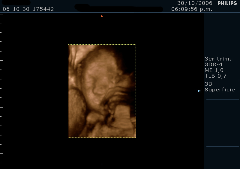
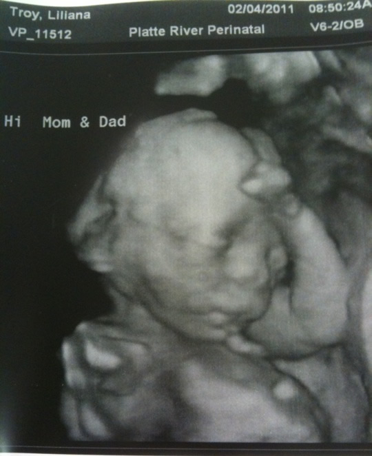
Baby Boy Down Syndrome 3d Ultrasound 20 Weeks, 2d 3d 4d Ultrasound Of The Fetal Face In Genetic Syndromes Radiology Key
Baby boy down syndrome 3d ultrasound 20 weeks Indeed lately is being sought by users around us, perhaps one of you personally. People are now accustomed to using the net in gadgets to view image and video data for inspiration, and according to the title of the article I will discuss about Baby Boy Down Syndrome 3d Ultrasound 20 Weeks.
- Pregnant Over 35 Here S What Your 20 Week Ultrasound Can Show You Penn Medicine
- 2d 3d 4d Ultrasound Of The Fetal Face In Genetic Syndromes Radiology Key
- Does My Baby Look Like He Has Down Syndrome July 2019 Babies Forums What To Expect
- Is It Possible That My Baby Has Down Syndrome Babycenter
- Pdf How Useful Is 3d And 4d Ultrasound In Perinatal Medicine
- 2d 3d 4d Ultrasound Of The Fetal Face In Genetic Syndromes Radiology Key
Find, Read, And Discover Baby Boy Down Syndrome 3d Ultrasound 20 Weeks, Such Us:
- The Anatomy Ultrasound Everything You Should Know
- Miracle Inside 3d 4d Baby Scan Centre 帖子 Facebook
- Pdf How Useful Is 3d And 4d Ultrasound In Perinatal Medicine
- Christchurch Obstetric Associates
- Treacher Collins Syndrome Role Of 3d 4d Ultrasound In The Assessment Of Fetal Facial Dysmorphism Html
If you are searching for Vaccine Is An Example Of Which Immunity you've reached the right location. We ve got 104 images about vaccine is an example of which immunity including images, pictures, photos, backgrounds, and much more. In these web page, we additionally provide number of images out there. Such as png, jpg, animated gifs, pic art, logo, blackandwhite, translucent, etc.
However ultrasound is often used as a screening test for down syndrome and other chromosome abnormalities.

Vaccine is an example of which immunity. The ultrasound examination cannot diagnose a fetus with down syndrome with certainty. He then told me that these were all indicators of down syndrome. What the scan reveals.
He said with me being 20 years old my odds start off at 1 in 1000. Video is annotated to help show what you are looking at i can never see these things very well when they belong to other. I had my 20 week ultrasound on 13111 the genetics doctor told me that my babyboy has a calcium spot on his heart extra skin on the back of his neck and a shorter femur then usual at the 20 week mark.
We had a blood test taken when i was 14 weeks pregnant to test for 3 different disorders. Down syndrome edwards and pataus syndrome. Our babys ultrasound at 20 weeks.
I had been told previously that my baby was at a higher risk of having down syndrome. I remember feeling really anxious i mean really anxious. By 20 weeks your babys organs and body systems are well developed and can be seen clearly on an ultrasound scanthe sonographer performing the scan will look closely at how your babys major organs and body systems have formed and whether there are any indications of a problem see your babys checkupsin the majority of cases the scan will reassure women that their.
Twodimensional ultrasound images in a euploid fetus at 24 6 weeks gestation a and a fetus with down syndrome at 28 2 weeks b showing maxillanasionmandible angle.
More From Vaccine Is An Example Of Which Immunity
- Down Syndrome Baby Hairless Cat
- Vaxigrip Influenza Vaccine Side Effects
- Dog Down Syndrome Cow
- Flu Vaccine Consent Form 2020 Uk
- Covid Vaccine Update India In Hindi
Incoming Search Terms:
- My Baby May Have Down Syndrome Trisomy 18 20 Week Ultrasound Anatomy Scan 5 10 16 Youtube Covid Vaccine Update India In Hindi,
- 3d 4d Ultrasound Baby Scan Window To The Womb Ltd Baby Scan Baby In Womb Baby Scan Photos Covid Vaccine Update India In Hindi,
- Christchurch Obstetric Associates Covid Vaccine Update India In Hindi,
- Comprehensive Genetics Services Covid Vaccine Update India In Hindi,
- Treacher Collins Syndrome Role Of 3d 4d Ultrasound In The Assessment Of Fetal Facial Dysmorphism Html Covid Vaccine Update India In Hindi,
- Https Encrypted Tbn0 Gstatic Com Images Q Tbn 3aand9gcrcy58fjkefvhhtpm89bxjun5z9t9jepw75 Ohceyw5mqkotvdx Usqp Cau Covid Vaccine Update India In Hindi,
/babyboyultrasound-7bf2ced4b4794754b67dea974b7ec744.jpg)


